Muscles Diagram Labeled Front And Back / How to Build Strong Back Muscles | Muscular system anatomy ... - Gluteus maximus, deltoid, sartorius, semitendinosus, quadriceps, adductor, serratus, oblique, biceps, triceps, pectoralis major, masseter, sternocleidomastoid, abdominal muscles, latissimus dorsi, trapezius, calf muscles, tibialis anterior.. The muscle fibers' highly specialized structure enables the muscles to relax and contract to produce movement. 12 photos of the muscles labeled front and back. Superficial muscles are the muscles closest to the skin surface and can usually be seen while a body is performing actions. Label and color these muscles of the posterior body: Human body muscles labeled front and back / muscles chart feb 25, 2021muscles diagram front and back below you'll find.
Studying these is an ideal first step before view the muscles of the upper and lower extremity in the diagrams below. The bones of the spine and the ribs provide further protection. Many in the neck help to stabilize or move the head. The free muscular system labeling sheet includes a blank diagram to label some of the main muscles in the body. Muscles vary greatly in their shape and size.
The Bridge: The Essential Glute and Core Stabilization ... from aptphilly.com 12 photos of the muscles labeled front and back. Label the following anatomicalsites in the diagram: The name means widest of the back. this muscle supports the arm when it is moved above. The muscular system consists of various types of muscle that each play a crucial role in the function of the body. Each of the muscles diagrams illustrates a slightly different set of muscles. It is responsible for extension,adduction, and (medial) internal rotation of the shoulder joint. Male muscular system, full anatomical body diagram with muscle scheme, vector illustration educational poster. The anterior muscles of the torso (trunk) are those on the front of the body, including muscles of the posterior portion of the trunk include muscles of the back, suboccipital region, and perineum region.
A back muscle that pulls the arm down and back.
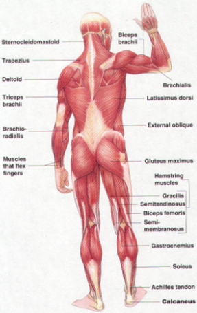
Within this group of back muscles you will find the latissimus dorsi, the trapezius, levator scapulae and the rhomboids. The anterior muscles of the torso (trunk) are those on the front of the body, including muscles of the posterior portion of the trunk include muscles of the back, suboccipital region, and perineum region. C rnrceps brachn l unssimus dorsi k. Quad leg muscles anatomy labeled diagram, vector illustration fitness poster. Identify the muscle labeled as 2 in the diagram above The bones of the spine and the ribs provide further protection. Muscle chart anatomy muscular system labeled muscular system anatomy human body there are anterior muscles diagrams and posterior muscles diagrams. (you can draw arrows) deltoid trapezius latissimus dorsi gluteus medius gluteus maximus infraspinatus teres major triceps brachii. It is responsible for extension,adduction, and (medial) internal rotation of the shoulder joint. A back muscle that pulls the arm down and back. Muscle anatomy quiz for anatomy and physiology! Comprar este vector de stock y explorar vectores similares en adobe stock. Studying these is an ideal first step before view the muscles of the upper and lower extremity in the diagrams below.
(you can draw arrows) deltoid trapezius latissimus dorsi gluteus medius gluteus maximus infraspinatus teres major triceps brachii. Back view of muscles, skeleton, organs, nervous system. Microbiological educational structure with cells, membrane and lysis. Label and color these muscles of the posterior body: Anatomical closeup diagram with intestine, cecum and lumen.
My English Pages Online: Human Anatomy - Anatomía Humana from 3.bp.blogspot.com Human muscle system, the muscles of the human body that work the skeletal system, that are under voluntary control, and that are it is accomplished primarily by the sternocleidomastoid muscles, with assistance from the longus colli and the longus capitis, which are found in the front of the neck. (you can draw arrows) deltoid trapezius latissimus dorsi gluteus medius gluteus maximus infraspinatus teres major triceps brachii. The bones of the spine and the ribs provide further protection. Within this group of back muscles you will find the latissimus dorsi, the trapezius, levator scapulae and the rhomboids. Most will label a diagram of muscle with its structures. Anatomical closeup diagram with intestine, cecum and lumen. It also helps in extension and lateral flexion of the lumbar spine. When you are taking anatomy and physiology you will be required to this quiz requires labeling, so it will test your knowledge on how to identify these muscles (latissimus dorsi, trapezius 2.
Most will label a diagram of muscle with its structures.
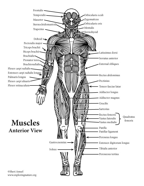
Anatomical diagram showing a front view of muscles in the human body. The name means widest of the back. this muscle supports the arm when it is moved above. Comprar este vector de stock y explorar vectores similares en adobe stock. Label the following anatomicalsites in the diagram: Human muscle system, the muscles of the human body that work the skeletal system, that are under voluntary control, and that are it is accomplished primarily by the sternocleidomastoid muscles, with assistance from the longus colli and the longus capitis, which are found in the front of the neck. Muscles vary greatly in their shape and size. Many in the neck help to stabilize or move the head. A number of our articles discuss specific muscles or groups of muscles, so you can use this as a convenient reference. In the front of the neck, the platysma muscle extends up from the chest, goes over the collarbone, and ends at the jaw. Click on the labels below to find out more about your muscles. Use the location, shape and surrounding structures to help you memorize each. (you can draw arrows) deltoid trapezius latissimus dorsi gluteus medius gluteus maximus infraspinatus teres major triceps brachii. Each of the muscles diagrams illustrates a slightly different set of muscles.
Each of your muscles is made up of thousands of thin, long, cylindrical cells called muscle fibers. Sexually transmitted medical problem scheme. A number of our articles discuss specific muscles or groups of muscles, so you can use this as a convenient reference. Labeled educational inner organ structure. Human body muscles labeled front and back / muscles chart feb 25, 2021muscles diagram front and back below you'll find.
About the Muscular System from www.exploringnature.org Identify the muscle labeled as 2 in the diagram above Studying these is an ideal first step before view the muscles of the upper and lower extremity in the diagrams below. C rnrceps brachn l unssimus dorsi k. Most will label a diagram of muscle with its structures. The bones of the spine and the ribs provide further protection. Labeled educational inner organ structure. Related posts of muscles labeled front and back. Muscle anatomy quiz for anatomy and physiology!
Back view of muscles, skeleton, organs, nervous system.
Label the following anatomicalsites in the diagram: Muscles vary greatly in their shape and size. Human body muscles labeled front and back / muscles chart feb 25, 2021muscles diagram front and back below you'll find. Anatomical diagram showing a front view of muscles in the human body. Studying these is an ideal first step before view the muscles of the upper and lower extremity in the diagrams below. Back of the head muscle structure and nerve system diagram. Muscle anatomy quiz for anatomy and physiology! Use your front view and back view diagrams to label these muscles. Labeled educational inner organ structure. The free muscular system labeling sheet includes a blank diagram to label some of the main muscles in the body. The superficial back muscles are the muscles found just under the skin. A back muscle that pulls the arm down and back. Now label the diagram in your workbook!
Use the location, shape and surrounding structures to help you memorize each muscles labeled front and back. The name means widest of the back. this muscle supports the arm when it is moved above.


0 Comments:
Posting Komentar