Home
Uncategories
Leg Bones Diagram / Leg Picture Image On Medicinenet Com : The bones of your leg have roughened patches on their surfaces where muscles are attached.
Leg Bones Diagram / Leg Picture Image On Medicinenet Com : The bones of your leg have roughened patches on their surfaces where muscles are attached.
Leg Bones Diagram / Leg Picture Image On Medicinenet Com : The bones of your leg have roughened patches on their surfaces where muscles are attached.. Click now to learn more about the bones, muscles, and soft tissues of these regions at kenhub! Arm bones (= bones in arm) are part of the appendicular skeleton which includes the hands, arms and shoulder girdle (clavicle and scapula) and the feet note: Lower jaw (mandible) collar bone. Visit kenhub for more skeletal system quizzes. We'll break down the anatomy and function of the upper leg, knee, lower leg, ankle, and foot.
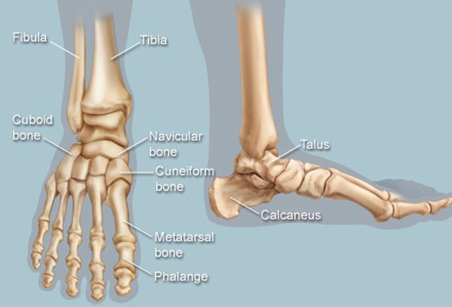
The bone that goes from your pelvis to your knee is called the femur (say: They are primarily compact bone but may have a large amount of spongy bone at the ends or extremities. The foot bones shown in this diagram are the talus, navicular, cuneiform, cuboid, metatarsals and calcaneus. This diagram shows the bones of the femur and the patella. Your leg bones are very large and strong to help support the weight of your body.
Leg Anatomy from fpnotebook.com The knee joint is the largest joint in the body and is primarily a hinge joint, although some sliding and rotation occur. The bones of your leg have roughened patches on their surfaces where muscles are attached. The femur, or thigh bone, is the largest, heaviest, and strongest bone in the human body. We'll break down the anatomy and function of the upper leg, knee, lower leg, ankle, and foot. At the distal end of the femur, two rounded condyles meet the tibia and fibula bones of the lower leg to form the knee joint. Visit kenhub for more skeletal system quizzes. Master leg and knee anatomy using our topic page. Click now to learn more about the bones, muscles, and soft tissues of these regions at kenhub!
You'll learn about the muscles, bones, and other structures of each area of the leg.
The humerus and the femur are corresponding bones of the arms and legs, respectively. It expands at the proximal and distal ends, articulating at the knee and ankle joints respectively. The foot bones shown in this diagram are the talus, navicular, cuneiform, cuboid, metatarsals and calcaneus. It mainly serves as an attachment point for the muscles of the lower leg. The bones of your leg have roughened patches on their surfaces where muscles are attached. The femur, or thigh bone, is the largest, heaviest, and strongest bone in the human body. Lower jaw (mandible) collar bone. While their parts are similar in general, their structure has been adapted to differing functions. At the microscopic level, this hard outer shell is made up of rod like structures called osteons. Want to learn more about it? The information above used to be on the page 'skeletal structures of the feet and hands' in the form of simple labelled diagrams of the leg. The bone that goes from your pelvis to your knee is called the femur (say: The knee joint is the largest joint in the body and is primarily a hinge joint, although.
Want to learn more about it? The information above used to be on the page 'skeletal structures of the feet and hands' in the form of simple labelled diagrams of the leg. Your leg bones are very large and strong to help support the weight of your body. The bones of your leg have roughened patches on their surfaces where muscles are attached. While their parts are similar in general, their structure has been adapted to differing functions.
File Human Arm Bones Diagram Svg Wikipedia from upload.wikimedia.org The bones of your leg have roughened patches on their surfaces where muscles are attached. Skeleton leg ankle joints and toe phalanges, cuboid, metatarsal, navicular and cuneiform bones, hand drawn dorsal view of foot. The knee joint is the largest joint in the body and is primarily a hinge the bones of the leg are the femur, tibia, fibula and patella.the foot bones shown in this diagram are the talus, navicular, cuneiform, cuboid. Long bones include bones of the thigh, leg, arm, and forearm. Human leg bones vector image. Want to learn more about it? Time to jump right into the biggest and strongest bones in the human body. Visit kenhub for more skeletal system quizzes.
File is ready to render.
Visit kenhub for more skeletal system quizzes. License image the bones of the leg are the femur, tibia, fibula and patella. Skeleton leg ankle joints and toe phalanges, cuboid, metatarsal, navicular and cuneiform bones, hand drawn dorsal view of foot. Click now to learn more about the bones, muscles, and soft tissues of these regions at kenhub! Includes leg (femur, tibia, patella, and fibula) and foot (tarsals and digits) bones. They are primarily compact bone but may have a large amount of spongy bone at the ends or extremities. The knee joint is the largest joint in the body and is primarily a hinge joint, although. The bones of the leg are the femur, tibia, fibula and patella. While their parts are similar in general, their structure has been adapted to differing functions. You'll learn about the muscles, bones, and other structures of each area of the leg. The humerus and the femur are corresponding bones of the arms and legs, respectively. Your legs are two of your most important body parts. At the distal end of the femur, two rounded condyles meet the tibia and fibula bones of the lower leg to form the knee joint.
When you stand or walk, all the weight of your upper body rests on them. Cheek bone (zygoma) upper jaw (maxilla). Skeleton leg ankle joints and toe phalanges, cuboid, metatarsal, navicular and cuneiform bones, hand drawn dorsal view of foot. File is ready to render. The foot bones shown in this diagram are the talus, navicular, cuneiform, cuboid, metatarsals and calcaneus.
Feet Human Anatomy Bones Tendons Ligaments And More from img.webmd.com Most bones (particularly the long bones of the arms and legs — which make up the appendicular skeleton) have a hard outer shell known as cortical bone. The foot bones shown in this diagram are the talus, navicular, cuneiform, cuboid, metatarsals and calcaneus. He leg's main function in the human is for locomotion and support of the rest of the body. When you stand or walk, all the weight of your upper body rests on them. Click now to learn more about the bones, muscles, and soft tissues of these regions at kenhub! At the microscopic level, this hard outer shell is made up of rod like structures called osteons. Your leg bones are very large and strong to help support the weight of your body. Includes obj for maximum compatibility.
Lower jaw (mandible) collar bone.
Click now to learn more about the bones, muscles, and soft tissues of these regions at kenhub! Learn vocabulary, terms and more with flashcards, games and other study tools. At the distal end of the femur, two rounded condyles meet the tibia and fibula bones of the lower leg to form the knee joint. Most bones (particularly the long bones of the arms and legs — which make up the appendicular skeleton) have a hard outer shell known as cortical bone. Time to jump right into the biggest and strongest bones in the human body. Master leg and knee anatomy using our topic page. When you stand or walk, all the weight of your upper body rests on them. The bones of your leg have roughened patches on their surfaces where muscles are attached. The tibia is the main bone of the leg, forming what is more commonly known as the shin. Your legs are two of your most important body parts. The knee joint is the largest joint in the body and is primarily a hinge the bones of the leg are the femur, tibia, fibula and patella.the foot bones shown in this diagram are the talus, navicular, cuneiform, cuboid. The foot bones shown in this diagram are the talus, navicular, cuneiform, cuboid, metatarsals and calcaneus. Its lower end helps create the knee joint.


0 Comments:
Posting Komentar